HRCT Chest (High-Resolution CT Scan)
Advanced Imaging for Detailed Evaluation of Lung Structure
What is an HRCT Chest?
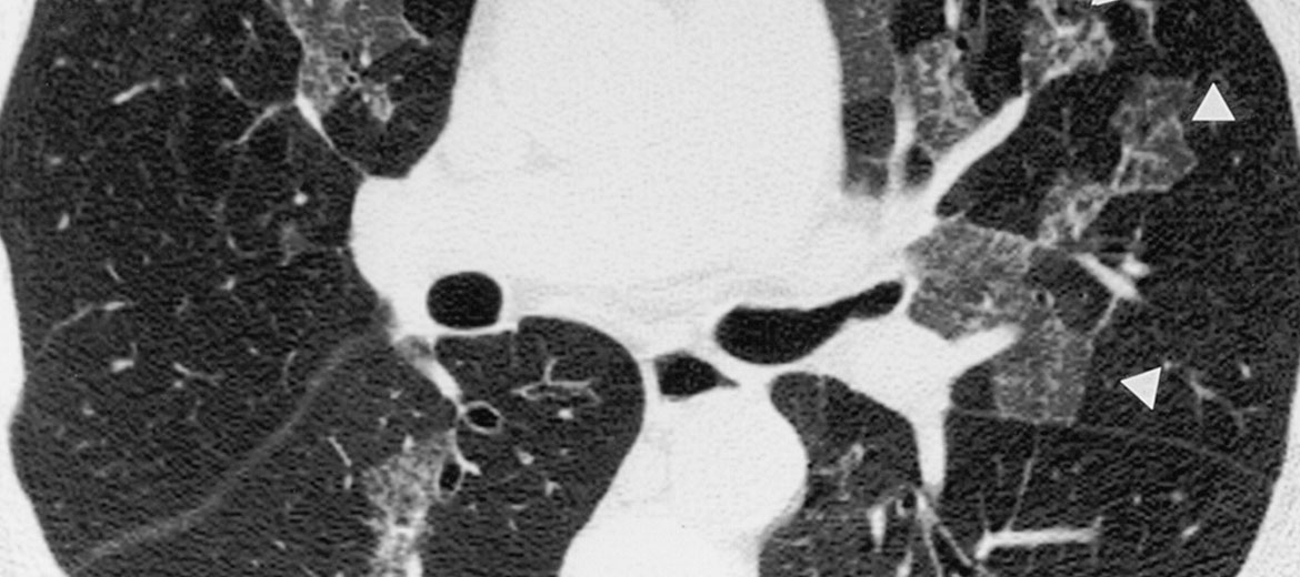
High-Resolution Computed Tomography (HRCT) of the chest is an advanced imaging test that provides detailed cross-sectional images of the lungs and surrounding structures.
Unlike a standard chest X-ray, HRCT offers superior resolution, allowing early detection of interstitial lung diseases, fibrosis, infections, bronchiectasis, and other complex lung conditions.
Why is an HRCT Chest Performed?
Who Should Consider an HRCT Chest?
Patients with persistent breathlessness, chronic cough, suspected interstitial lung disease, abnormal chest X-ray findings, recurrent infections, or suspected lung fibrosis may require HRCT imaging. It is also recommended for detailed evaluation of lung damage after severe infections or inflammatory conditions.
How is the HRCT Chest Performed?
The patient lies comfortably on a scanning table that moves through a CT scanner. The machine captures high-resolution images of the lungs in thin slices.
The procedure is painless and usually completed within 10–15 minutes. In some cases, contrast dye may be used for better visualization, depending on the clinical requirement.